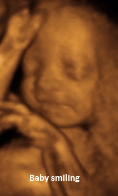
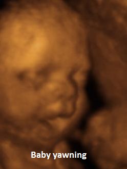
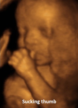
3D/4D Ultrasound Scans


Ultrasound is the only non-invasive way to see a baby in the uterus. Advances in imaging have resulted in the introduction of three-dimensional (3D) and four-dimensional (4D) ultrasound. Used in conjunction with traditional two-dimensional (2D) ultrasound, 3D/4D ultrasound provides more detail in “real” time, so your baby can be seen moving around.
A 3D/4D ultrasound scan can be undertaken at any stage in pregnancy but the best time is after 26 weeks for the most realistic pictures. At this stage providing your baby is lying in the correct position we will clearly be able to see your baby’s face, hands, fingers, feet and toes as well as any facial movements or gestures it may be making.
There can be limitations sometimes with a 3D/4D scan. Things that make it difficult to see the baby clearly include inadequate amniotic fluid surrounding the baby, its face covered by its hands and feet or if it is looking away making it difficult to get a clear picture.
Safety of Ultrasound
Ultrasound is an imaging technique that has been used routinely in ante natal screening for the last 20 years and there are no known effects of it when used responsibly by qualified Ultrasonographers and within the guidelines laid down by the British Medical Ultrasound Society. Therefore in order for us to practice safely and responsibly, the Ultrasonographers at KMI will only carry out a 4D scan in conjunction with one of the other routine scans offered.
In some cases 3D/4D will not possible and whilst every effort will be made to obtain good pictures, the scan time cannot be extended indefinitely and we may need to rebook you for a different day.
What is included with this scan?
We will give you a report, pictures, and a CD with all your pictures on if requested (in Jpeg format) to take home with you.
Recording on a mobile phone
We are happy for you to do some recording on your mobile phone, but we ask that you keep this for personal use and do not share it on social media. The ultrasonographer will look at your baby before you start recording to check that it has a heartbeat and that there are no major problems. None of the ultrasonographers will provide a running commentary, they will stay quiet whilst you are recording, and we request that you do not video the ultrasonographer undertaking your examination.


